- Mammography Guidelines for Women with Breast Implants
- Mammography Guidelines for Following Breast Reduction Surgery
- Magnetic Resonance (MR) Imaging of Breast Implants
Women with breast implants should follow the same American Cancer Society (ACS) program of recommended mammograms as women without breast implants (click here to view the ACS guidelines). However, due to the implant, several special mammography views must be taken to allow visualization of both the breast tissue and the implant. For this reason, diagnostic mammography is usually performed on patients with breast implants (as opposed to screening mammography that is typically performed on asymptomatic women without implants).
Examination of the augmented breasts is more time consuming; therefore, the imaging location performing the mammography should be informed of the presence of implants when the mammogram is scheduled. Patients with implants should also inform the physician and the technologist performing the exam that they have implants. Imaginis.com is unaware of any documented cases where mammography has been the direct cause of implant rupture.
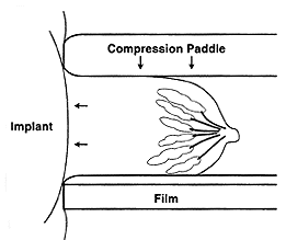
The x-rays used for mammographic imaging of the breasts cannot penetrate silicone or saline implants well enough to image the overlying or underlying breast tissue. Therefore, some breast tissue (approximately 25%) will not be seen on the mammogram, as it will be covered up by the implant. In order to visualize as much breast tissue as possible, women with implants undergo four additional views as well as the four standard images taken during diagnostic mammography. In these additional x-ray pictures, called Eklund views or implant displacement (ID) views, the implant is pushed back against the chest wall and the breast is pulled forward over it. This allows better imaging of the forward most part of each breast. The implant displacement views are not as successful in women who have contractures (formation of hard scar tissue around the implants). The ID views are easiest to obtain in a women whose implants are placed underneath (behind) the chest muscle.
 |
 |
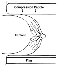
| Standard mammography
views are taken first. The breast and implant are compressed with moderate force |
Image displacement mammography views (also called Eklund views) are performed with the implant pushed back against the chest wall. The compression paddle is applied to the breast tissue, which is pulled forward. |
Women who have had breast contouring orbreast reduction (also called mastopexy or reduction mammaplasty) should also receive annual mammograms once they reach 40 years of age. It is important for the radiologist to be aware of the patient's surgical history. This will help the radiologist when they interpret the mammogram images.


