


Breast ultrasound can image several different types of breast conditions, including both benign (non-cancerous) and malignant (cancerous) lesions. Ultrasound is frequently used to evaluate breast abnormalities that are found with screening mammography or diagnostic mammography or during a physician performed clinical breast exam. Ultrasound allows significant freedom in obtaining images of the breast from almost any orientation. This section provides sample images of a variety of breast conditions that can be imaged with ultrasound. For general information on breast ultrasound, click here.
Sharp diagnostic ultrasound images are often able to show soft layers of breast tissue. Ultrasound may be particularly useful in detecting abnormalities in patients with dense breasts. Density is a term used to describe breast tissue that has many glands close together. Though fairly common (especially in younger women), dense breasts may make breast masses difficult to detect on a mammogram film.
All images are courtesy of Siemens Medical Solutions.
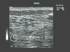
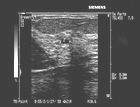
This ultrasound image shows the subtle differentiation of glandular tissue in a normal breast.
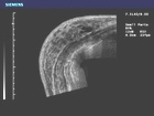
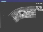
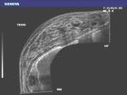
This detailed image of normal breast tissue uses panoramic ultrasound imaging.
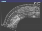
This detailed image was taken after a tumor was removed.
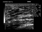
This image shows superficial breast ducts.
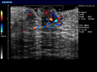
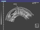
With color visualization, this image shows the small superficial vessels of the nipple.
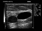
Breast cysts, tiny accumulations of fluid, are the most common cause of benign (non-cancerous) breast lumps in women between age 30 and 50. Simple cysts are typically round or oval and have smooth edges. Complex cysts can be filled with debris and may sometimes require aspiration to confirm that they are indeed benign cysts. Both single and multiple cysts are very common. The exact causes of cysts are not known, but they do tend to change with hormonal variations, either during normal menstrual cycles or from post-menopausal hormone replacement therapy.
When examining a breast abnormality to determine whether it is a cyst (or multiple cysts), the radiologist will study the quantity, size, and internal characteristics of the abnormality. Cysts do not become cancer or increase the risk of cancer. Most of the time, cysts may be left alone, but sometimes a physician may drain them with a small needle using ultrasound guidance. This procedure is called cyst aspiration.
Breast cyst. Image shows sharp cystic walls and internal septation by cyst membrane.
Breast cyst. Image shows subtle contents within cyst.
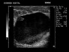
Large cyst with layered debris and a solid component.
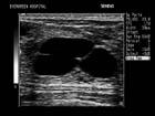
Adjacent breast masses: one a debris-filled cyst, the other a simple cyst.
Panoramic view of a simple breast cyst within the glandular layer of breast tissue.
This image shows atypical characteristics of this cystic breast structure: a somewhat thickened cystic wall and a 5-millimeter septation by cystic membrane.
Patient with multiple cysts and other masses that indicate breast disease. Ultrasound shows the relationship of the dominant cyst to other masses in this patient.
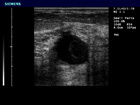
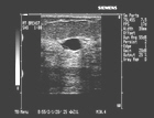
One application of breast ultrasound involves differentiating simple cysts from solid masses. This image shows a small cyst within the glandular breast tissue.
Panoramic image shows ductal and muscular layers of this high-density breast in addition to identifying multiple small cysts.
