


Nuclear medicine studies were first performed in the 1950s using special devices called "gamma cameras." Nuclear medicine studies require the oral or intravenous introduction of very low-level radioactive chemicals (called radionuclides, radiopharmaceuticals or radiotracers) into the body. Radiopharmaceuticals are specially formulated to be collected temporarily in the specific part of the body to be studied. The radionuclides are taken up by the organs in the body and then emit faint gamma ray signals which are measured by a gamma camera. The gamma camera has a large crystal detector (called a scintillation crystal). These crystals detect the emitted radiation signal and convert that signal into faint light. The light is then converted to an electric signal, which is then digitized (converted into a computer signal) and reconstructed into an image by a computer. The resulting image is viewed on the system monitor and can be manipulated (post-processed) and filmed, sent over a network to another location, or saved on a disk.
The nuclear medicine image can either be in grayscale (shades of black and white), for instance in a bone scan, or they can be color coded to clearly show functional activity, like in a cardiac study.
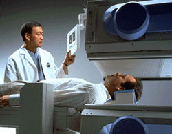
The technologist positions the patient and begins a "dual head" nuclear medicine examination. The devices above and below the patient are the dual gamma cameras and each contains a scintillation crystal and other image acquisition electronics.
In an x-ray or CT examination, the radiation comes out of the x-ray or CT system and then passes through the patient's body before being detected and recorded onto film or by a computer. Nuclear medicine uses the opposite approach: a radioactive material is introduced into the patient, and is then detected by a machine called a gamma camera. The radiation which is emitted by the body during nuclear medicine imaging are gamma rays. These gamma rays are similar to x-rays but have a shorter wavelength.
The radionuclide substances used in nuclear medicine imaging are usually either synthesized radioactive substances, like technetium, or radioactive forms of elements that are naturally found in the body, such as iodine. The levels of radiation involved in nuclear medicine studies is usually considerably lower than a patient would receive in a conventional x-ray study or CT scan.
Modern nuclear medicine equipment provides all digital (computerized) creation of the images. This means that it is possible for the nuclear medicine images to be:
- conveniently stored on various archive media or in multiple locations
- networked or sent to other locations within an imaging center or around the world for additional professional interpretation or consultation or
- combined with other digital patient medical record data like patient history, other exams and imaging studies, and therapy records.
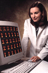
The radiologist reviews a bone scan using the nuclear medicine computer workstation
The net result of the nuclear medicine information being available digitally is that best patient care can be made quickly and cost effectively, with few restrictions on location, time of day, or type of procedure.
Updated: June 10, 2008
