Evaluating a Breast Lump Return to Previous

The discovery of a breast lump—whether by chance, during a routine breast self-exam, or during a clinical breast exam—can be stressful for a woman. Because a lump can be a symptom of breast cancer, all persistent breast lumps should be evaluated by a physician. However, the majority of breast lumps (approximately 80%) are due to non-cancerous causes.

Main Menu:

What Should a Woman Do If She Finds a Breast Lump?

First, it is important for all women to practice monthly breast-self exams beginning at age 20. These self-exams allow women to become familiar with how their breasts look and feel so they can more readily detect any changes that may occur. Many women naturally have some lumpiness and asymmetry (differences between the right and left breast). The key to the breast self-exam is to learn to find changes in the breast(s) that persist over time. If a new lump is found and does not disappear after the menstrual cycle, then it should be reported to a physician for clinical evaluation.


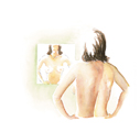
All persistent breast lumps should be evaluated by a physician. Practicing monthly breast self-exams helps women get to know their breasts and more easily detect changes.

  Click here to learn more about breast self-exam.

What Signs Suggest a Lump is Likely to Be Cancerous?

It is not possible for a woman or a physician to know for certain whether a breast lump indicates breast cancer until imaging exams (such as mammography and ultrasound) and/or biopsy are performed. A breast biopsy involves taking a sample of breast tissue and examining it under a microscope to determine whether it contains cancer cells. However, there are certain characteristics associated with lumps that can suggest whether they are more likely to be cancer or benign (non-cancerous).

Signs that suggest a lump is more likely to be cancerous:

  • The lump is firm and hard
  • The lump is not discrete; it is not easily distinguishable
  • The lump is fixed in the breast; it does not move
  • There is only one lump
  • There is not an identical lump in the opposite breast
  • The skin of breast is dimpled
  • The lump is accompanied by bloody nipple discharge

Signs that suggest a lump is less likely to be cancerous:

  • The lump is soft
  • The lump is discrete; it is easily distinguishable
  • The lump moves in the breast
  • There are multiple breast lumps
  • There is an identical lump in the opposite breast
  • The lump disappears after the menstrual cycle

While the above signs can help suggest whether a lump is more likely or less likely to be cancerous, having one or more of these characteristics does not guarantee or eliminate the possibility of having breast cancer. These characteristics merely provide clues for the physician when evaluating a lump. Some breast cancers can have characteristics found in the "less likely to be cancerous" category. Therefore, all persistent breast lumps need to be presented to a physician.

Fibrocystic breasts: Fibrocystic breast condition is a common, non-cancerous condition that affects more than 50% of women at some point in their lives. In fact, the condition is so common that many physicians refrain from using the term "fibrocystic" and simply tell their patients that their breasts are lumpier than average but are still normal.

The most common signs of fibrocystic breasts include: lumpiness, tenderness, cysts, areas of thickening, fibrosis, and breast pain. Having fibrocystic breasts, in and of itself, is not a risk factor for breast cancer. However, fibrocystic breast condition can sometimes make it more difficult to detect a hidden breast cancer with standard examination and imaging techniques. Therefore, it is important that women with fibrocystic breasts practice monthly breast self-exams, receive regular clinical breast exams, and have yearly screening mammograms (the latter beginning at age 40).

Symptoms of Fibrocystic Breasts

  • cysts (fluid-filled sacs)
  • fibrosis (scar-like connective tissue)
  • lumpiness
  • areas of thickening
  • tenderness
  • pain

The degree to which women experience symptoms of fibrocystic breast condition varies considerably. Some women with fibrocystic breasts have only mild breast pain and may not be able to feel any breast lumps when performing breast self-exams. Other women with fibrocystic breasts may experience more severe breast pain or tenderness and may feel multiple lumps in their breasts. Most fibrocystic breast lumps are found in the upper, outer quadrant of the breasts (near the armpit), although these lumps can occur anywhere in the breasts. Fibrocystic breast lumps tend to be smooth, rounded, and mobile (not attached to other breast tissue), though some fibrocystic tissue may have a thickened, irregular feel. The lumps or irregularities associated with fibrocystic breasts are often tender to touch and may increase or decrease in size during the menstrual cycle.

  Click here to learn more about fibrocystic breasts.

How Are Breast Lumps Evaluated By Physicians?

Whether a breast lump is first detected by a physician during a clinical breast exam or by the woman herself, the process of evaluation usually begins with a detailed patient history. The physician will ask the patient specific questions about the lump and her medical history to help identify the cause of the lump. Sample questions may include:

  • How long have you had the lump?
  • Does the lump change in size with your menstrual cycle?
  • How long has it been since your last menstrual period?
  • Have you recently been pregnant or are you breast-feeding?
  • Have you experienced discharge from the nipple?
  • Do you use hormone replacement therapy?
  • Have you experienced any recent trauma to the breast?
  • Have you had any previous breast biopsies? If yes, what were the diagnoses?
  • Do you have a history of cancer?
  • Do you have a history of other medical conditions?
  • Have you had a mammogram or other breast imaging test before?

These questions can provide important information as to what is causing the lump. For example, a woman who has recently been pregnant and who is breast-feeding may have a galactocele (milk-filled cyst). A woman who is taking hormone replacement therapy (HRT) may have more nodules in her breast due to the therapy. Trauma to the breast may cause a hematoma (a blood-filled packet), fat necrosis (swelling of fatty breast tissue), or a ruptured cyst (fluid-filled packet).

Learning the patient’s family and personal medical history can also be helpful. A family history of breast cancer can increase a woman’s chances of developing breast cancer herself. A personal history of non-cancerous conditions such as atypical hyperplasia (an abnormal increase in breast cells) or lobular carcinoma in situ (LCIS) can also increase the risk of breast cancer.

Once a thorough patient history is taken, the physician will perform a thorough clinical breast exam to investigate the lump and other areas of the breast and axilla (armpit). In addition to feeling for breast masses, the physician will check for any skin dimpling, nipple retraction, or other visual changes. The clinical breast exam typically lasts several minutes and the patient will usually need to raise her arms, place her hands on her hips and exert pressure, and lie down during the exam so the breasts can be examined from different angles.

After the clinical breast exam, the evaluation of a breast lump will differ depending on the woman’s age, history, and characteristics of the lump. The following descriptions provide information on how women in different age groups are typically evaluated:

  • Women age 30 or older: A diagnostic mammogram is usually ordered. A diagnostic mammogram differs from the routine screening mammogram in that it involves additional x-ray views from different angles and/or special magnification. A diagnostic mammogram is used instead of a screening mammogram when a breast abnormality is present. Depending on the results of the mammogram, additional breast imaging (such as ultrasound) may be ordered. In many cases, further breast imaging will be ordered even if a mammogram does not show a suspicious abnormality. This is because a small percentage of breast cancers can be missed with mammography. Depending on the results of the mammogram and additional imaging tests, a breast biopsy may be performed. A biopsy involves removing a sample of breast tissue and examining it under a microscope to determine whether cancer cells are present. Sometimes, a biopsy (or fine needle aspiration—sampling of a few breast cells) will be performed even if breast imaging tests are normal. This usually happens when the physician suspects that the breast lump is suspicious regardless of the results of the imaging tests.
  • Women under age 30: In this group of women, a mammogram may or may not be the first test ordered. This is because mammography is not always beneficial in younger women who tend to have dense breast tissue which can mask breast cancer and other abnormalities on a mammogram film. In some cases, ultrasound or other tests may be performed. However, mammography can still be beneficial in some women younger than 30. If the breast imaging tests reveal a suspicious abnormality, a biopsy may be ordered to examine a sample of breast tissue. As with women over age 30, a biopsy (or fine needle aspiration—sampling a few breast cells) may be performed even if breast imaging tests are normal. Again, this usually happens when the physician suspects that the breast lump is suspicious regardless of the results of the imaging tests.

In approximately 80% of cases, breast lumps are benign (non-cancerous). Benign conditions that can cause breast lumps include:

  • Fibrocystic breasts
  • Cysts
  • Fibroadenomas
  • Papillomas
  • Phyllodes tumors (usually benign)
  • Galactoceles
  • Granular cell tumors
  • Duct ectasia
  • Fat necrosis

  Click here to learn more about these benign conditions.

If a biopsy reveals breast cancer, then the woman and her cancer team will discuss treatment options. Treatment options include surgery (lumpectomy or mastectomy), radiation, chemotherapy, and/or other drug therapies, such as tamoxifen. Practicing monthly breast self exams, receiving regular clinical breast exams, and yearly screening mammograms (the latter beginning at age 40) can help detect breast cancer early when the chances of successful treatment and survival are the greatest.

 Click here to learn more about treating breast cancer. 

Breast Lumps During Pregnancy

Breast cancer during pregnancy can be difficult to diagnose because the breasts naturally undergo several changes. During pregnancy, the breasts increase in size and become more tender, especially during the first half of pregnancy. The most rapid period of breast growth is during the first eight weeks of pregnancy. As the pregnancy progresses, the breasts become firmer and more nodular to prepare for lactation (breast-feeding). It is very important for women to continue to perform monthly breast self-exams during pregnancy and receive monthly physician-performed clinical breast exams so as not to delay the possible diagnosis of breast cancer.

As in non-pregnant women, the majority of breast lumps found during pregnancy are benign (non-cancerous). However, because a lump can signal breast cancer, all persistent lumps should be evaluated by a physician. Approximately one in 3,000 (0.03%) to one in 10,000 (0.01%) women are diagnosed with breast cancer during pregnancy. Breast cancer itself does not appear to harm a fetus.

If a lump is detected during pregnancy, an ultrasound exam and/or mammogram will typically be performed. Ultrasound is excellent at distinguishing cysts (packets of fluid) and is routinely used for fetal imaging because it does not harm the fetus. Mammography is also considered safe for pregnant women and the fetus because it uses a very low dose of radiation. In many cases, a biopsy will be performed if a suspicious breast lump is detected in a pregnant woman. A biopsy confirms or denies the presence of breast cancer.

Non-cancerous conditions that are common during pregnancy include:

  • Cysts (collections of fluid)
  • Galactoceles (milk-filled cysts)
  • Fibroadenomas (tumors; existing ones may enlarge during pregnancy)

If breast cancer is detected during pregnancy, it is not necessary to terminate the pregnancy. Treatment options should be discussed with the patient’s cancer team. Surgery, such as lumpectomy and mastectomy, can be performed safely during pregnancy. Radiation, chemotherapy, and drug therapies (such as tamoxifen) are usually delayed until after childbirth.

  Click here to learn more about pregnancy and breast cancer.

Additional Resources and References

Updated: May 4, 2008

Click here to learn how to perform breast self-exam

Return to the Breast Health Main Menu

This Article Can Be Found At:

Click Here to Recommend This Article To A Friend

Home | About Us | Free Newsletter | News | Bookstore | Return to Previous


Imaginis.com subscribes to the HONcode principles of the Health on the Net Foundation.

Copyright © 1997-2008 Imaginis Corporation. All rights reserved. Imaginis® is a registered service mark of Imaginis Corporation, a health services company. All other trademarks and copyrights are property of their respective owners. Note: Use of this online service is subject to the disclaimer and the terms and conditions contained herein. Information found within the Imaginis.com website or in other sites linked to from Imaginis.com is provided for educational purposes only and is not intended to be used as a substitute for diagnosis and treatment by a medical doctor. Imaginis does not endorse and has no responsibility for the content of any other sites listed on Imaginis.com, and provides links and references merely as a convenience to its users. Seek immediate medical attention if your condition is urgent.