


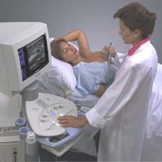
Breast ultrasound, also known as sonography or ultrasonography, is frequently used to evaluate breast abnormalities that are found with screening or diagnostic mammography or during a physician performed clinical breast exam. Ultrasound allows significant freedom in obtaining images of the breast from almost any orientation. Ultrasound is excellent at imaging cysts: round, fluid-filled, pockets inside the breast. Additionally, ultrasound can often quickly determine if a suspicious area is in fact a cyst (always non-cancerous) or an increased density of solid tissue (dense mass) which may require a biopsy to determine if it is malignant (cancerous).
If a patient's ultrasound and mammogram results are both negative (no evidence of cancer is seen), but the physician is still concerned about the thickening or mass, then he/she may proceed further with a fine needle aspiration biopsy (FNA) of the area.
Though breast ultrasound has excellent contrast resolution, it lacks the detail (spatial resolution) of conventional mammography, and therefore, ultrasound is not approved by the U.S. Food and Drug Administration (FDA) as a screening tool for breast cancer. Rather, ultrasound is used to investigate an abnormality detected by mammography or during a physician performed breast exam. Currently, only mammography is FDA approved to look for breast cancer in asymptomatic women (women with no signs or symptoms of breast cancer).
Physicians use ultrasound to evaluate breast abnormalities that have been found with screening or diagnostic mammography or during a clinical breast exam. Ultrasound may help detect some breast masses and is the best way to determine whether a cyst is present without placing a needle into the area of concern to aspirate fluid. Ultrasound is also useful in helping physicians guide a biopsy (tissue sampling) to determine whether a breast abnormality is cancerous. Physicians use ultrasound during core and fine needle aspiration biopsies (FNA) to determine where to place the needle. Ultrasound may also be used to prove whether a suspicious area is a lymph node. Lymph nodes have fatty centers which are often apparent on ultrasound images.
Ultrasound has excellent contrast resolution. This means, for example, that an area of fluid (cyst) and an area of normal breast tissue are easy to differentiate on an ultrasound image. However, ultrasound does not have good spatial resolution like mammography, and therefore cannot provide as much detail as a mammogram image. Ultrasound is also unable to image microcalcifications, tiny calcium deposits that are often the first indication of breast cancer. Mammography, on the other hand, is excellent at imaging calcifications. Ultrasound may be able to detect macrocalcifications (larger calcium deposits) in some cases.
Though most true breast lumps will be found by mammography or ultrasound, some abnormalities escape detection on both imaging tests. For example, a lump may be able to be felt but does not appear on mammography or ultrasound images. If this is the case, then fine needle aspiration biopsy (FNA) is often performed. Less than 30% of all breast biopsies are cancerous. In cases where the abnormality is not apparent on mammogram or ultrasound, the chances of cancer are significantly less.
