


- Steps to reduce risk of stroke and heart attack
- Surgery of the carotid arteries to help prevent stroke (carotid endarterectomy)
Not having a stroke in the first place is vastly better than relying on medical science to undo the damage of stroke after it has been done. Keeping cholesterol levels down decreases the risk of atherosclerosis and blood clots, both major causes of stroke. Treating high blood pressure drastically reduces the risk of bleeding (hemorrhagic) strokes. Eliminating smoking also improves a person's chances of not having a stroke. Exercise, proper diet and elimination of smoking are among the best prescriptions to minimize the health conditions that cause stroke and heart disease.
|
|
|
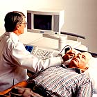
| The
ultrasongrapher positions the transducer on the patients neck to image the right carotid.
Image courtsey Siemens Medical Systems, Inc. |
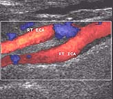
A color
ultrasound showing the right carotid arteries. Normal blood flow is in red. Obstructed
blood flow (due to stenosis) is shown as blue and can be seen at the bifurcation
(branching) of the carotid artery.
Image courtsey Siemens Medical Systems, Inc. |
Medical care for patients at high risk of stroke may include the administration of one adult aspirin daily (to thin the blood) and aggressive management of modifiable risk factors, such as counseling to help them stop smoking and treatment for high blood pressure, high cholesterol, and diabetes when indicated. These treatments may involve the use of drugs to lower cholesterol and blood pressure.
- reduce weight
- control high blood pressure
- reduce sodium
- quit smoking
- exercise regularly
- control cholesterol
- eat less fat
- reduce stress
Carotid artery surgery (carotid endarterectomy) can prevent stroke in carefully selected individuals who have no outward sign of disease but are at risk for stroke from a severe narrowing of the carotid artery in the neck. A carotid endarterectomy is a surgical procedure in which a vascular surgeon or neurosurgeon removes fatty deposits and plaque from one of the two main arteries in the neck supplying blood to the brain.
Carotid artery disease is more common in elderly people. The disease process that causes the buildup of fat and other material on the artery walls is called atherosclerosis, popularly known as "hardening of the arteries." Narrowing of an artery is called stenosisand the degree of stenosis is usually expressed as a percentage of the normal diameter of the opening. Carotid stenosis can be measured using high resolution ultrasound, magnetic resonance angiography, CT angiography and other techniques. Click here to learn about the use of medical imaging to diagnose vascular disease before a stroke occurs.
In 1992, the most recent year for which statistics are available from the National Hospital Discharge Survey, there were about 91,000 carotid endarterectomies performed in the United States. The average cost of carotid endarterectomy including diagnostic test, surgical procedure, hospitalization, and follow-up care is about $15,000.
Two large clinical trials have identified specific individuals for whom the carotid surgery is highly beneficial when performed by surgeons and in institutions that can match the standards set in those studies. Carotid surgery has been found highly beneficial for persons who have already had a stroke or experienced the warning signs of a stroke and have a severe stenosis of 70 percent to 99 percent. In this group, surgery reduces the estimated two-year risk of stroke by more than 80 percent, from greater than 1 in 4 to less than 1 in 10. In a second trial, the carotid surgery has also been found highly beneficial for persons who are have no symptoms but have a severe carotid stenosis of 60 percent to 99 percent. In this group, the surgery reduces the estimated 5-year risk of stroke by more than one-half, from about 1 in 10 to less than 1 in 20.
Other methods of treating carotid artery disease include the use of angioplasty and stenting to open the stenosis (narrowing). Stenting i
