


The early detection of ovarian cancer greatly increases the chances that the disease can be successfully treated. If patients are diagnosed with ovarian cancer before the cancer has spread outside the ovary, nine out of ten women typically survive five years or longer. However, the American Cancer Society estimates that only 25% of ovarian cancer cases are diagnosed in early stages. This is because the symptoms of ovarian cancer are often subtle and not recognized until the disease has progressed to advanced stages.
If a patient experiences symptoms of ovarian cancer or an abnormality is detected during a pelvic exam, a number of imaging tests may be ordered to evaluate the situation. If imaging tests warrant further evaluation, a pelvic biopsy may be performed to confirm whether or not the patient has ovarian cancer.
Ultrasound/sonogram: Ultrasound uses sound waves to create clear pictures of the ovaries on a video monitor. Either an abdominal or transvaginal ultrasound exam may be performed to examine the ovaries. An abdominal ultrasound involves moving a hand-held transducer over the abdomen to create images of the ovaries. A transvaginal ultrasound involves inserting an ultrasound probe, slightly larger than a tampon, into the vagina to obtain images of the ovaries. Researchers are investigating whether transvaginal ultrasound may be effective at screening women who are at high risk of ovarian cancer but who do not have any symptoms of the disease.
 |
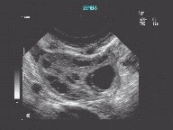 |
| Color flow ultrasound imaging showing normal ovarian flow. | Ultrasound image showing ovarian follicles (pockets). |
CT scan: Also called CAT scan (computed tomography), this exam combines the use of a digital computer together with a rotating x-ray device to create detailed cross sectional images of an organ or body part.
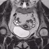
MRI scan: Also called MR scan (magnetic resonance), this exam uses magnetic energy and radio waves to create cross-sectional images of an organ or body part.
 |
 |
| A soft "pelvis coil" is positioned on the patient just prior to MR imaging of the pelvis and ovaries |
Coronal MR image of the female pelvis showing the ovaries and uterus |
 |
 |
| High resolution coronal MR image of the female pelvis clearly shows
both ovaries.
Image courtesy of Glenn Coates, MD, Raleigh MRI/Wake Radiology (www.wakerad.com). |
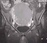
Coronal
MR image showing large ovarian cystadenocarcinoma. Gadolinium contrast enhances the frond
like projections from the cyst wall.
Image courtesy of Glenn Coates, MD, Raleigh MRI/Wake Radiology (www.wakerad.com). |
Blood test: In some instances, a sample of blood may be analyzed to determine its level of CA-125 (also called OC-125), a tumor marker for ovarian cancer. However, this test is not always an accurate method of determining whether a woman has ovarian cancer and is usually performed in conjunction with other tests.
If imaging tests suggest that a woman may have ovarian cancer, a biopsy may be performed to confirm the presence of cancer. A biopsy involves removing a portion of the ovary tissue (or fluid) and analyzing it under a microscope to determine whether it contains cancer cells. Tissue samples are obtained during surgery (often a laparotomy or laparascopy).
Laparotomy: This is a surgical procedure that involves an incision in the lower abdomen between the navel and the pubic area. Essentially, a laparotomy is exploratory surgery; the surgeon examines the area and removes a sample of fluid or tissue for analysis. If the surgeon suspects that cancer is present during the operation, he or she may go ahead and remove the entire ovary during surgery. This helps prevent the spread of cancer cells into the abdominal cavity that could occur when only the outer layer of the ovary is cut during biopsy.
Laparascopy: This surgical procedure involves passing a flexible, lighted tube through a small incision in the abdomen. As with a laparotomy, the surgeon examines the area and removes a sample of fluid or tissue for analysis. Again, the entire ovary may be removed, if necessary, to prevent the further spread of cancer.
If cancer is confirmed with microscopic analysis, the patient and her cancer team should begin weighing treatment options. Click here to learn how ovarian cancer is treated.
Updated: January 10, 2008
