


Fetal-ultrasound (also called prenatal ultrasound) has become an almost automatic part of the child birth process during visits to the obstetrician. Ultrasound is routinely used at 16 to 18 weeks to date the pregnancy and check on the development of the fetus.
The tremendous feelings a mother has for her child growing inside her womb are hard to describe. The experience is different, yet wonderful, for every mother. The sensation many mothers and fathers feel when they first glimpse live ultrasound images of their fetal babies bring a fascinating reality and a new dimension to the parental experience of pregnancy and child birth.
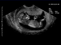
Prenatal ultrasound showing baby's head, body, arms and legs
The ultrasonic waves used to image the fetus (or other organs) cannot be heard and produce no sensation to the person (or fetus) being imaged. A key benefit of ultrasound is that it uses no x-ray and is safe for both the fetus and the mother. Some women may have one to three ultrasound examinations during the course of a normal pregnancy.
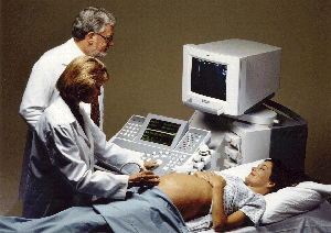
Mother, doctor and ultrasound technologist experience the joy of prenatal ultrasound
Modern obstetric medicine (for guiding pregnancy and child birth) relies heavily on ultrasound to provide detailed images of the fetus and uterus.
Fetal Ultrasound can show:
- the fetal baby's physical development and even determine age of pregnancy
- functions like breathing, heartbeat, elimination, and body movement
- images of limbs and spinal column to check for proper formation and growth
- development of the brain and other major organs
- whether there is multiple pregnancy (twins, triplets, ...)
Ultrasound is also used to guide prenatal surgery such as the safe delivery of medication to the fetus inside the womb.
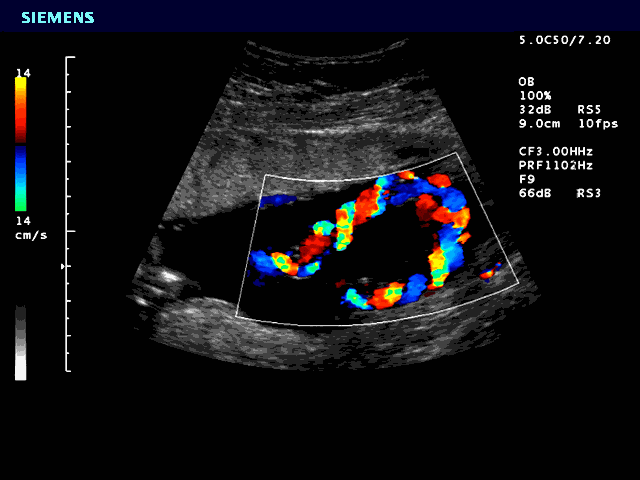
Ultrasound of umbilical cord, color shows blood supply to the fetus
Fetal ultrasound is a straightforward process that is quick and painless, a typical exam only takes about 15 minutes. The mother is usually examined while lying on her back. The clothing over the abdomen and pelvis region is removed and a jelly like solution is applied to the skin. This gel helps improve the contact between the ultrasound transducer and the skin, allowing better images of the fetus.
During the acquisition of images, the ultrasound transducer is passed back and forth over the area of interest. In-audible, high frequency sound waves are emitted by the ultrasound transducer and as these sound waves pass into the body, they are reflected back at different strengths and frequencies by the different anatomic structures of both the mother and the fetus. The same transducer receives these reflected sound waves and a computer reconstructs them into an image in real time. The obstetrician, the ultrasound technologist and the mother can watch this beautiful movie unfold before their eyes.
During the examination, the ultrasound "movie" can often be recorded to video tape or the movie can be frozen (like viewing a video tape in "freeze frame" mode) and a still image can be recorded onto paper or film. Typically, moving ultrasound images (images being acquired and displayed at multiple frames per second) give a much better appreciation of the structure of the fetus or other anatomy being imaged than a still image. A new, state-of-the-art development in ultrasound is the ability to scan a panoramic view. As the transducer is moved along a part of the body, the image continues to grow, showing the entire scanned area (instead of just showing the area currently being scanned by the transducer). This "panoramic landscape view" gives the obstetrician an appreciation for both fine detail and the "big picture."
