


- Ultrasound Scanning
- Digital Imaging Techniques
- Benefits of digital technology to all x-ray systems:
- Computed Tomography (CT)
- Magnetic Resonance (MR)
Radiology began as a medical sub-specialty in first decade of the 1900's after the discovery of x-rays by Professor Roentgen. The development of radiology grew at a good pace until World War II. Extensive use of x-ray imaging during the second world war, and the advent of the digital computer and new imaging modalities like ultrasound and magnetic resonance imaging have combined to create an explosion of diagnostic imaging techniques in the past 25 years.
Milestones in Medical Diagnosis and Diagnostic Imaging
For the first fifty years of radiology, the primary examination involved creating an image by focusing x-rays through the body part of interest and directly onto a single piece of film inside a special cassette. In the earliest days, a head x-ray coul d require up to 11 minutes of exposure time. Now, modern x-rays images are made in milliseconds and the x-ray dose currently used is as little as 2% of what was used for that 11 minute head exam 100 years ago. Further, modern x-ray techniques (both anal og film screen systems and digital systems, described below) have significantly more spatial resolution and contrast detail. This improved image quality allows the diagnosis of smaller pathology that could not be detected with older technology.
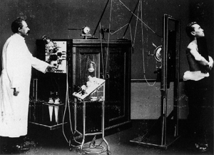
An x-ray system from the pioneering days.
Patients still had to hold the cassettes themselves.
The next development involved the use fluorescent screens and special glasses so the doctor could see x-ray images in real time. This caused the doctor to stare directly into the x-ray beam, creating unwanted exposure to radiation. In 1946, George Sc hoenander developed the film cassette changer which allowed a series of cassettes to be exposed at a movie frame rate of 1.5 cassettes per second. By 1953, this technique had been improved to allow frame rates up to 6 frames per second by using a special "cut film changer."
A major development along the way was the application of pharmaceutical contrast medium to help visualize organs and blood vessels with more clarity and image contrast. These contrast media agents (liquids also referred to as "dye") were fir st administered orally or via vascular injection between 1906 and 1912 and allowed doctors to see the blood vessels, digestive and gastro-intestinal systems, bile ducts and gall bladder for the first time.
In 1955, the x-ray image intensifier (also called I.I.) was developed and allowed the pick up and display of the x-ray movie using a TV (television) camera and monitor. By the 1960's, the fluorescent system (which had become quite complex with mirror optic systems to minimize patient and radiologist dose) was largely replaced by the image intensifier/TV combination. Together with the cut-film changer, the image Intensifier opened the way for a new radiologic sub-specialty know as angiography to bloss om and allowed the routine imaging of blood vessels and the heart.
Nuclear Medicine studies (also called radionuclide scanning) were first done in the 1950s using special gamma cameras. Nuclear medicine studies require the introduction of very low-level radioactive chemicals into the body. These radionuclides are taken up by the organs in the body and then emit faint radiation signals which are measured or detected by the gamma camera.
In the 1960's the principals of sonar (developed extensively during the second world war) were applied to diagnostic imaging. The process involves placing a small device called a transducer, against the skin of the patient near the region of interest, for example, the kidneys. This transducer produces a stream of inaudible, high frequency sound waves which penetrate into the body and bounce off the organs inside. The transducer detects sound waves as they bounce off or echo back from the internal st ructures and contours of the organs. These waves are received by the ultrasound machine and turned into live pictures with the use of computers and reconstruction software.
Digital imaging techniques were implemented in the 1970's with the first clinical use and acceptance of the Computed Tomography or CT scanner, invented by Godfrey Hounsfield. Analog to digital converters and computers were also adapted to conventional fluoroscopic image intensifier/TV systems in the 70's as well. Angiographic procedures for looking at the blood vessels in the brain, kidneys, arms and legs, and the blood vessels of the heart all have benefited tremendously from the adaptation of digital technology.
Over the next ten to fifteen years a large majority of conventional x-ray systems will also be upgraded to all digital technology. Eventually, all of the film cassette/film screen systems will be replaced by digital x-ray detectors. This technology i s currently works-in-progress and is only available at a handful of sites worldwide. An intermediate step called phosphor plate technology in currently available at hundreds of sites around the world. These plates trap the x-ray energy and require an in termediate processing step to release the stored information so it can be converted into a digital picture.
- less x-ray dose can often be used to achieve the same high quality picture as with film
- digital x-ray images can be enhanced and manipulated with computers
- digital images can be sent via network to other workstations and computer monitors so that many people can share the information and assist in the diagnosis
- digital images can be archived onto compact optical disk or digital tape drives saving tremendously on storage space and manpower needed for a traditional x-ray film library
- digital images can be retrieved from an archive at any point in the future for reference.
Some modalities like mammography require extremely high resolution film to show the small breast cancers. Digital detectors capable of a similarly high resolution are under development and will hopefully be available in the future. However, digital imag ing is already being used in parallel to high resolution film in breast imaging and breast biopsy systems.
CT imaging (also called CAT scanning for Computed Axial Tomography) was invented in 1972 by Godfrey Hounsfield in England. Hounsfield used gamma rays (and later x-rays) and a detector mounted on a special rotating frame together with a digital compute r to create detailed cross sectional images of objects. Hounsfield's original CT scan took hours to acquire a single slice of image data and more than 24 hours to reconstruct this data into a single image. Today's state-of-the-art CT systems can acquire a single image in less than a second and reconstruct the image instantly.
The invention of CT was made possible by the digital computer. The basic algorithms involved in CT image reconstruction are based on theories proposed by the scientist Radon in the late 1700's. To honor his remarkable discovery, Hounsfield was awarde d the Nobel Prize and was granted Knighthood by the Royal Family of England.
An original head-only CT scanner from 1974
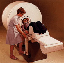
MR principals were initially investigated in the 1950s showing that different materials resonated at different magnetic field strengths. Magnetic Resonance (MR) Imaging (also know as MRI) was initially researched in the early 1970s and the first MR im aging prototypes were tested on clinical patients in 1980. MR imaging was cleared for commercial, clinical availability by the Food and Drug Administration (FDA) in 1984 and its use throughout the U.S. has spread rapidly since.
Countless scientists have been involved in the innovation of magnetic resonance. The development of MR imaging is attributed to Paul Lauterbur and scientists at Thorn-EMI Laboratories, England, and Nottingham University, England.
Updated: December 30, 2008
