


What is Endoscopy and Why is it Performed?
Endoscopy allows physicians to peer through the body's passageways. Endoscopy is the examination and inspection of the interior of body organs, joints or cavities through an endoscope. An endoscope is a device that uses fiber optics and powerful lens systems to provide lighting and visualization of the interior of a joint. The portion of the endoscope inserted into the body may be rigid or flexible, depending upon the medical procedure.
An endoscope uses two fiber optic lines. A "light fiber" carries light into the body cavity and an "image fiber" carries the image of the body cavity back to the physician's viewing lens. There is also a separate port to allow for administration of drugs, suction, and irrigation. This port may also be used to introduce small folding instruments such as forceps, scissors, brushes, snares and baskets for tissue excision (removal), sampling, or other diagnostic and therapeutic work. Endoscopes may be used in conjunction with a camera or video recorder to document images of the inside of the joint or chronicle an endoscopic procedure. New endoscopes have digital capabilities for manipulating and enhancing the video images.
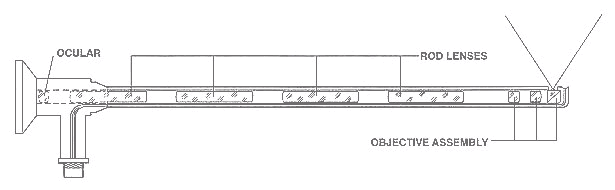
This figure shows a rigid endoscope used for arthroscopy. The "image fiber" leads from the ocular (eye piece) to the inserted end of the scope. The "light fiber" is below and leads from the light source to the working end of the endoscope.
Why Is Endoscopy Performed?
Endoscopy can be used to diagnose various conditions by close examination of internal organ and body structures. Endoscopy can also guide therapy and repair, such as the removal of torn cartilage from the bearing surfaces of a joint. Biopsy (tissue sampling for pathologic testing) may also be performed under endoscopic guidance. Local or general anesthetic may be used during endoscopy, depending upon the type of procedure being performed.
Internal abnormalities revealed through endoscopy include: abscesses, biliary (liver) cirrhosis, bleeding, bronchitis, cancer, cysts, degenerative disease, gallbladder stones, hernia, inflammation, metastatic cancer, polyps, tumors, ulcers, and other diseases and conditions.
Endoscopy is a minimally invasive procedure and carries with it certain minor risks depending upon the type of procedure being performed. However, these risks are typically far outweighed by the diagnostic and therapeutic potential of the procedure.
Prior to the widespread use of endoscopy and diagnostic imaging, most internal conditions could only be diagnosed or treated with open surgery. Until the last several decades, exploratory surgery was routinely performed only when a patient was critically ill and the source of illness was not known. For example, in certain dire cases, the patient's thorax or abdomen were surgically opened and examined to try to determine the source of illness.
Endoscopy can often be done on an outpatient basis. "Outpatient" means that the procedure does not require hospital admission and acute care and observation and may be performed outside the premises of a hospital. Outpatient procedures performed at hospitals or ambulatory centers allow the patient to go home or return to work within a short while after their procedure.
Types of Endoscopy
Fiber optic endoscopes now have widespread use in medicine and guide a myriad of diagnostic and therapeutic procedures including:
- Arthroscopy: examination of joints for diagnosis and treatment (arthroscopic surgery)
- Bronchoscopy: examination of the trachea and lung's bronchial trees to reveal abscesses, bronchitis, carcinoma, tumors, tuberculosis, alveolitis, infection, inflammation
- Colonoscopy: examination of the inside of the colon and large intestine to detect polyps, tumors, ulceration, inflammation, colitis diverticula, Chrohn's disease, and discovery and removal of foreign bodies.
- Colposcopy: direct visualization of the vagina and cervix to detect cancer, inflammation, and other conditions.
- Cystoscopy: examination of the bladder, urethra, urinary tract, uteral orifices, and prostate (men) with insertion of the endoscope through the urethra.
- ERCP (endoscopic retrograde cholangio-pancreatography) uses endoscopic guidance to place a catheter for x-ray fluorosocopy with contrast enhancement. This technique is used to examine the liver's biliary tree, the gallbladder, the pancreatic duct and other anatomy to check for stones, other obstructions and disease. X-ray contrast is introduced into these ducts via catheter and fluoroscopic x-ray images are taken to show any abnormality or blockage. If disease is detected, it can sometimes be treated at the same time or biopsy can be performed to test for cancer or other pathology. ERCP can detect biliary cirrhosis, cancer of the bile ducts, pancreatic cysts, pseudocysts, pancreatic tumors, chronic pancreatitis and other conditions such as gallbladder stones.
- EGD (Esophogealgastroduodensoscopy): visual examination of the upper gastro-intestinal (GI) tract. (also referred to as gastroscopy) to reveal hemorrhage, hiatal hernia, inflammation of the esophagus, gastric ulcers.
- Endoscopic biopsy is the removal of tissue specimens for pathologic examination and analysis.
- Gastroscopy: examination of the lining of the esophagus, stomach, and duodenum. Gastroscopy is often used to diagnose ulcers and other sources of bleeding and to guide biopsy of suspect GI cancers.
- Laparoscopy: visualization of the stomach, liver and other abdominal organs including the female reproductive organs, for example, the fallopian tubes.
- Laryngoscopy: examination of the larynx (voice box).
- Proctoscopy, sigmoidoscopy, proctosigmoidoscopy: examination of the rectum and sigmoid colon.
- Thoracoscopy: examination of the pleura (sac that covers the lungs), pleural spaces, mediastinum, and pericardium.
A Brief History of Endoscopy
In the early 1900s, the first attempts to view inside the body with lighted telescopes were made. These initial devices were often fully rigid. In the 1930s, semi-flexible endoscopes called gastroscopes were developed to view inside of the stomach. Fiber-optic endoscopy was pioneered by South African-born physician Basil Hirschowitz at the University of Michigan in 1957. Widespread use of fiber optic endoscopes began in the 1960s.
A fiber optic cable is simply a bundle of microscopic glass or plastic fibers that literally allows light and images to be transmitted through curved structures. Fiber optic cables are also replacing metal wires as the backbone of the world's telecommunications infrastructure. This Internet page may have traveled through a fiber optic cable as a stream of digital data (bursts of light) on its way to your computer.
