


Computed Tomography is based on the x-ray principal: as x-rays pass through the body, they are absorbed or attenuated (weakened) at differing levels creating a matrix or profile of x-ray beams of different strength. This x-ray profile is registered on film, thus creating an image. In the case of CT, the film is replaced by a banana shaped detector which measures the x-ray profile.
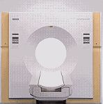
Outside view of modern CT system showing the patient table and CT scanning patient aperture
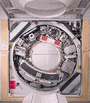
Inside view of modern CT system, the x-ray tube is on the top at the 1 o'clock position and the arc-shaped CT detector is on the bottom at the 7 o'clock position. The frame holding the x-ray tube and detector rotate around the patient as the data is gathered.
A CT scanner looks like a big, square doughnut. The patient aperture (opening) is 60 cm to 70 cm (24" to 28") in diameter. Inside the covers of the CT scanner is a rotating frame which has an x-ray tube mounted on one side and the banana shaped detector mounted on the opposite side. A fan beam of x-ray is created as the rotating frame spins the x-ray tube and detector around the patient (see figure below). Each time the x-ray tube and detector make a 360° rotation, an image or "slice" has been acquired. This "slice" is collimated (focused) to a thickness between 1 mm and 10 mm using lead shutters in front of the x-ray tube and x-ray detector.
As the x-ray tube and detector make this 360° rotation, the detector takes numerous snapshots (called profiles) of the attenuated x-ray beam. Typically, in one 360° lap, about 1,000 profiles are sampled. Each profile is subdivided spatially (divided into partitions) by the detectors and fed into about 700 individual channels. Each profile is then backwards reconstructed (or "back projected") by a dedicated computer into a two-dimensional image of the "slice" that was scanned.
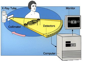
Diagram showing relationship of x-ray tube, patient, detector, and image reconstruction computer and display monitor
Multiple computers are used to control the entire CT system. The main computer that orchestrates the operation of the entire system is called the "host computer." There is also a dedicated computer that reconstructs the "raw CT data" into an image. A workstation with a mouse, keyboard and other dedicated controls allows the technologist to control and monitor the exam. The CT gantry and table have multiple microprocessors that control the rotation of the gantry, movement of the table (up/down and in/out), tilting of the gantry for angled images, and other functions such as turning the x-ray beam on an off.
