


 |
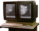
| The GE workstation allows "soft-copy" interpretation of digital mammography images. Image courtesy of GE Medical Systems |
An advisory panel to the U.S. Food and Drug Administration (FDA) has recommended the approval of the General Electric Medical Systems "full-field" digital mammography scanner to screen for and diagnose breast cancer. If approved by the FDA, digital mammography may eventually replace traditional mammography. The advantages of digital mammography include faster image acquisition, shorter exam, easier image storage, and physician manipulation of breast images for more accurate detection of breast cancer.
Digital (computerized) mammography is similar to standard screening mammography in that x-rays are used to produce detailed images of the breast. Full-field digital mammography uses essentially the same mammography system as conventional mammography, but the system is equipped with a digital receptor and a computer instead of a film cassette. Presently, only "spot-view" digital systems are FDA approved for use in guiding breast biopsy . Some digital spot-view systems can perform both cassette/film mammography and digital spot mammography.
Before applying for FDA certification, GE gathered data from 662 patients at four institutions: the University of Colorado, the University of Massachusetts Medical Center, Massachusetts General Hospital, and the Hospital of the University of Pennsylvania. The data compared hard copies of digital breast images on film to conventional mammography films finding that digital mammography is as effective at detecting breast cancer as standard film mammograms. A separate study revealed that the digital mammography scanner showed a slight advantage in the visibility of breast tissue at the skin line.
After reviewing the data, the FDA advisory panel voted unanimously that the digital mammography scanner is safe and effective. However, the panel recommended that GE continue to study the effectiveness of the digital mammography scanner in "soft-copy" applications and present those findings to the FDA when ready. Soft copy images are digital images that are interpreted on a computer monitor. Such soft-copy interpretation allows radiologists to use a high-resolution monitor and make adjustments for contrast and light intensity to better view the images.
With soft-copy reporting of digital mammogram images, magnification, orientation, brightness, and contrast of the breast image can be changed after the exam is completed to help the doctor more clearly see certain areas. Digital mammography also has the potential to significantly reduce the amount of time required to acquire a mammogram from 10 to 15 minutes to less than a minute. This will provide a shorter, more comfortable exam for the woman and possibly allow mammography facilities to conduct more mammograms in a day.
If the FDA accepts the advisory panel’s recommendation to approve the GE digital mammography scanner (known as the Senographe 2000D), system users will only be able to read digitally processed hard copies (films). Additional clinical trials are being conducted for soft-copy reporting. Digital mammography scanners are already used in Germany and Canada.
Standard mammography using film cassettes has the benefit of providing very high detail resolution (sharpness), which is especially useful for imaging microcalcifications (tiny calcium deposits) and very small abnormalities that may indicate breast cancer. While full field digital mammography may lack the spatial resolution of film, clinical trials showed the GE digital mammography system to be at least equivalent to standard film screening mammography. This is because digital mammography has the benefit of providing improved contrast resolution, which may make abnormalities easier to see. Fischer Imaging, Fuji Medical, Siemens Medical Systems, and Trex, are trying to develop digital mammography systems with detail resolution equivalent to standard film mammography while also providing the benefits of digital mammography noted above.
Many radiologists support digital mammography as an effective tool to screen for breast cancer. In an open letter to the FDA published in the December 1999 issue of Diagnostic Imaging, Dr. Etta Pisano, Chief of Breast Imaging and Professor of Radiology at the University of North Carolina-Chapel Hill, encouraged the FDA to quicken their methods of evaluating digital mammography. "We believe that digital mammography has advantages over standard screen-film mammography that are readily apparent from the study of phantom images and the physical parameters that can be measured. The contrast resolution of these devices is inherently better," said Dr. Pisano.
"In addition, the extra features that digital mammography will ultimately provide, such as telemammography, tomosynthesis, and computer-aided diagnosis will prove invaluable to patients and their doctors," said Dr. Pisano. Telemammography allows radiologists to share digital images via phone or network connection for remote consultation with other physicians; tomosynthesis allows radiologists to add or subtract digital mammography images using a computer workstation for enhanced diagnostic capability. Computer-assisted detection (CAD) was approved by the FDA in June 1999. CAD helps radiologists more accurately detect breast cancer by marking suspicious areas on digitized mammograms.
Though digital mammography promises to help improve breast cancer detection in the future, the system costs roughly four to five times as much as standard mammography equipment. Standard mammography systems are currently installed in over 10,000 locations across the United States. It may take years for this current equipment to be updated or replaced and for digital mammography to become widespread after its approval by the FDA.\
- The December 17, 1999 WebMD report, "FDA Panel Backs First Digital Mammography System," is available at http://my.webmd.com/content/article/1728.53497
- The December 17, 1999 Diagnostic Imaging report, "FDA Advisory Panel Gives GE Medical Green Light for Digital Mammography," is available at http://www.dimag.com/1999121701.asp
- The December 17, 1999 Associated Press report, "Digital Mammography a Step Closer," is reprinted on Excite at http://news.excite.com/news/ap/991217/11/digital-mammograms
- To learn more about digital mammography, please visit http://www.imaginis.com/breasthealth/digital_mammo.asp
