


In a stress thallium study, two data acquisitions are performed during a stress thallium study. First, the patient is vigorously exercised on a treadmill or stationary bicycle to elevate cardiovascular activity and "stress" the heart. This is followed immediately by a nuclear medicine examination. The patient is then given a period of time to rest. When the patient's heart activity has again become normal (or "at rest"), a second nuclear medicine study is completed. The physician then compares the images and function of the heart at rest to the heart under stress. Areas of the heart which may have been previously damaged by myocardial infarction (heart attack) or may have insufficient blood supply due to a blockage of a coronary artery will not show the proper function in the stress image. Another common cardiac application of nuclear medicine is the MUGA scan (electrocardiographic MUltiple Gated Acquisition) which allows study of the heart's muscular wall motion and study of the heart's chambers.
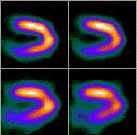
Cardiac "stress thallium" nuclear medicine study comparing the heart under stress (upper row) to the heart at rest (lower row). Study shows normal cardiac function.
Nuclear Medicine Can Image and/or Show the Function a Variety of Organs and Body Parts to Diagnose a Number of Medical Conditions Including:
- abdomen (example given, to check for gastrointestinal bleeding)
- brain (e.g., to look for tumors or aneurysms (blood vessel disease) or evaluate stroke)
- blood (e.g., to test for various blood cell disorders)
- breast (e.g., to image breast cancers)
- hepatobiliary system (e.g., to check gallbladder and bile duct function)
- heart (e.g., to look for coronary artery disease, myocardial infarction, valve disease or heart attack; to detect heart transplant rejection; check the effectiveness of bypass surgery; to select patients for angioplasty or bypass surgery)
- kidneys (e.g., to check renal function; to detect renal tumors; to test for renal transplant rejection)
- liver/spleen (e.g., to check for chirrhosis or metastatic cancer)
- lung (e.g., to check for pulmonary embolism (blood clot), check for lung transplant rejection or test for smoke inhalation injury in burn patients)
- lymphatic system (e.g., to detect if cancer has spread to the lymph nodes)
- skeletal system (e.g., to check for metastatic cancer or to test for hidden bone trauma in sports injuries)
- stomach (e.g., to check for stomach function and to confirm ulcers or cancer)
- thyroid and parathyroid (e.g., to check for tumor or abnormal function)
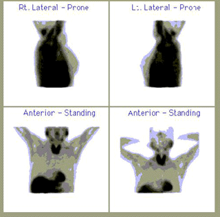
This nuclear medicine study shows normal breast anatomy and no abnormalities in this patient.
Updated: June 10, 2008
